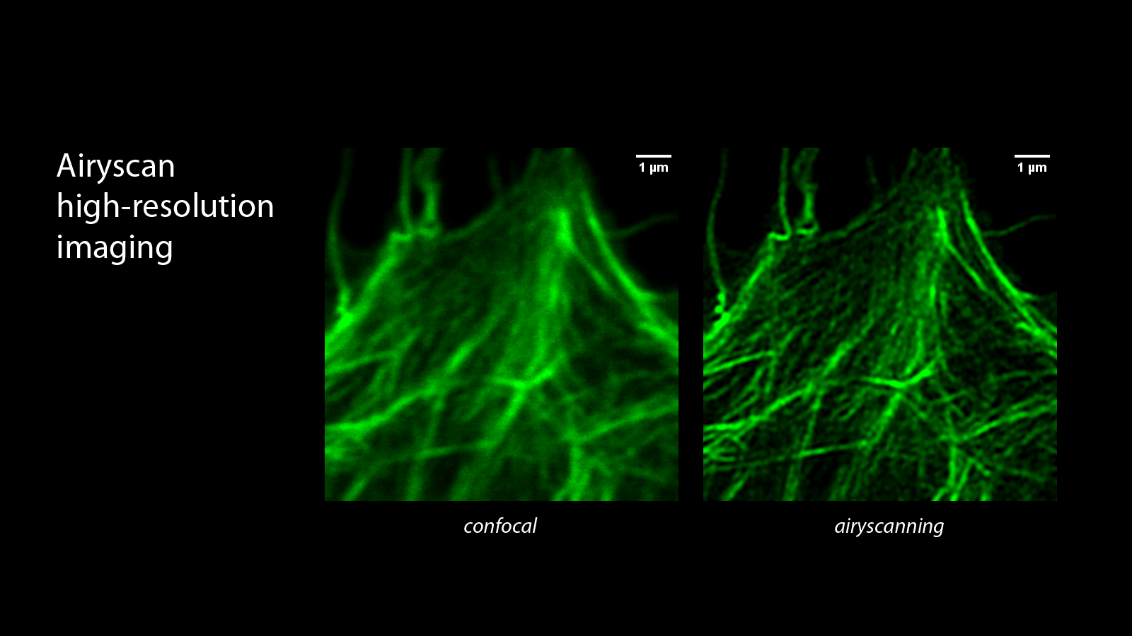
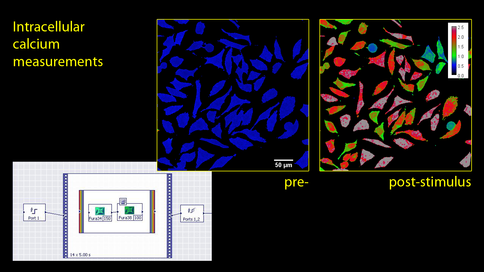
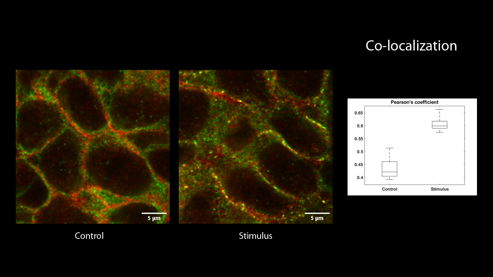
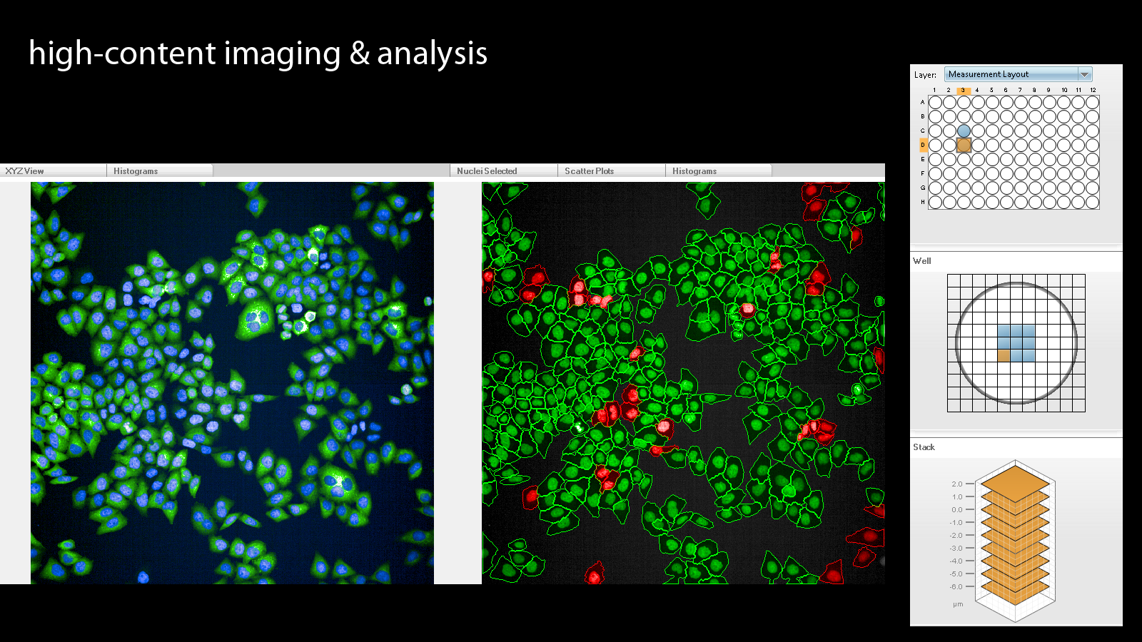
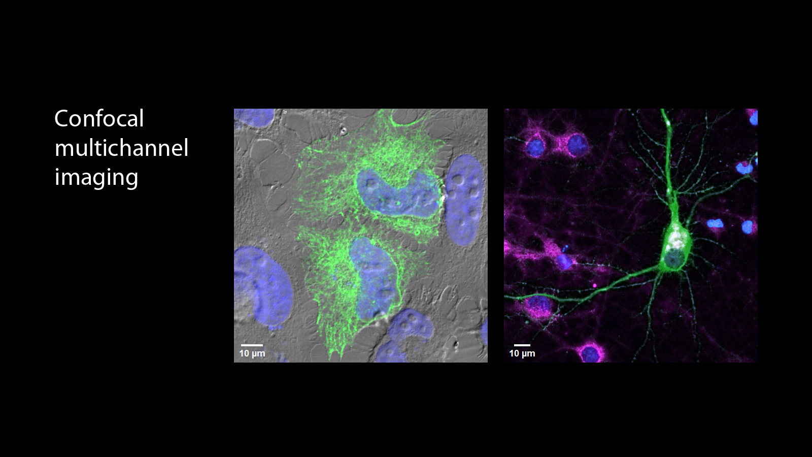
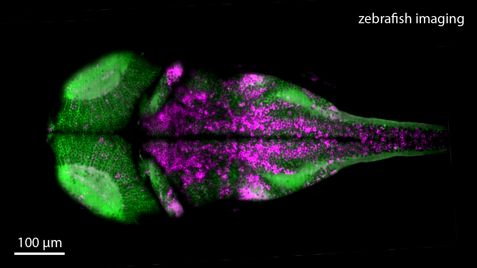
The facility provides access to a broad range of fluorescence light microscopes. Most of our microscopes allow optical sectioning (point-scanning confocal, spinning-disk confocal, two-photon, lightsheet, and total internal reflection fluorescence microscopy) to facilitate high-contrast fluorescence imaging. Typical applications include multichannel confocal imaging of cells and tissues, live-imaging of cells (on inverted stands with temperature and atmosphere control), live imaging of zebrafish larvae (on Zeiss Lightsheet Z.1), airyscan high-resolution imaging of cells (on Zeiss LSM 800), intracellular calcium imaging with Fura-2 (on Olympus CellR), and high-content imaging and analysis of cells and other biological specimens (PerkinElmer Opera Phenix). A more detailed description of the equipment is provided here.
The MCF Light Microscopy service is free of charge for internal researchers. External users from academia and industry are charged according to the pricelist.
For more information, please contact Tomasz Wegierski (twegierski@iimcb.gov.pl).
We offer:
Equipment access
- Prior training is required for most microscopes
Full service
(Only for external users and Opera Phenix high-content screening system)
- Consultation, design of experiments
- Image acquisition
- Image processing & analysis
- Preparation of figures
Note: we do not offer cell culturing and staining
